X-ray Tomography & Interferometry
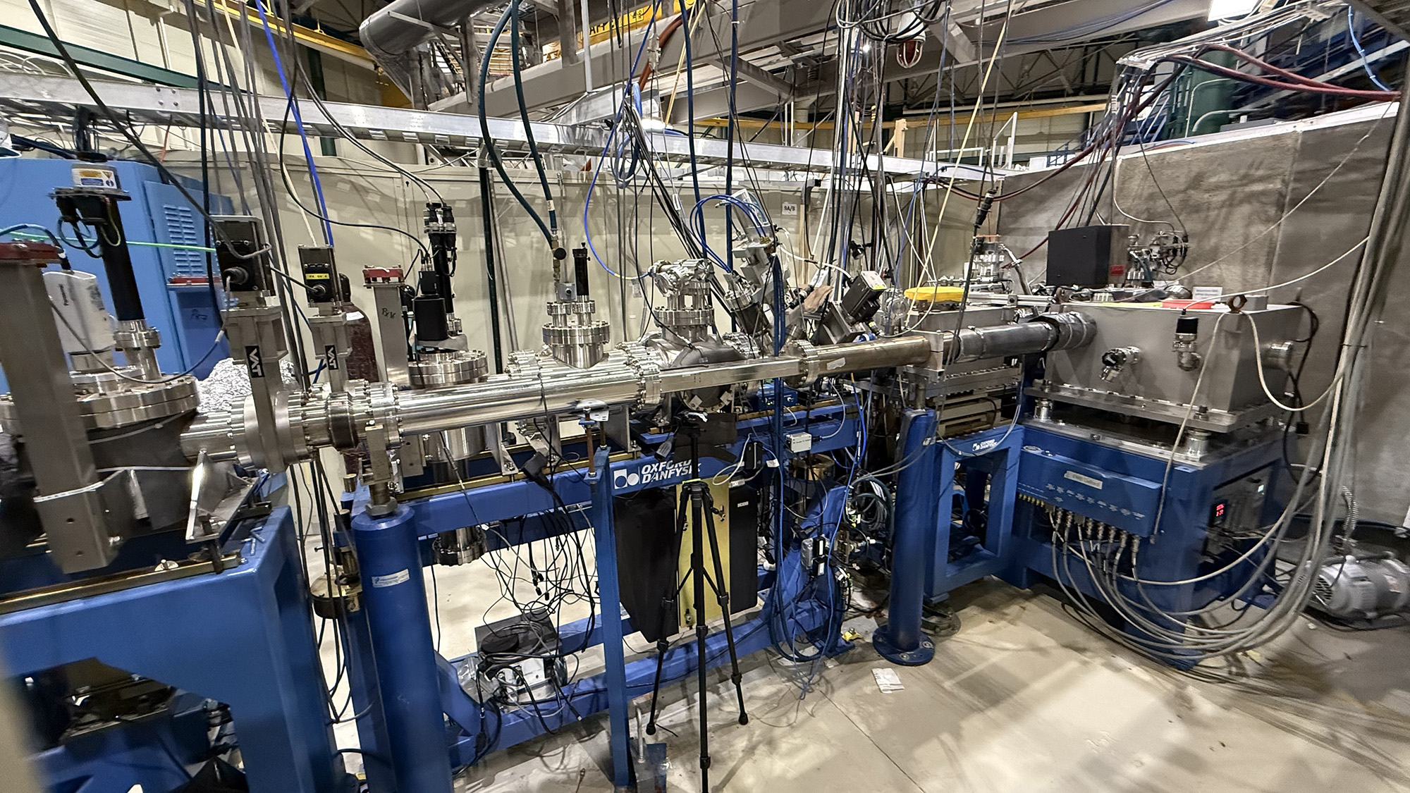
Tomography beamline
Tomography is 3D imaging of objects from a shadow cast by a beam. We use monochromatic X-rays up to 60 keV with an effective spatial resolution of 3 µm. The field of view is determined by the X-ray beam height (1-3 mm) and the pixel number in the sCMOS camera (2k x 2k). Thus, we can image a maximum volume of 3 mm x 5 mm x 5 mm at 3 µm resolution. Thus, the object should have a diameter smaller than 5 mm. The speed of tomography imaging ranges from one to two hours, depending upon camera exposure time (up to 10 seconds) and number of projections used to create the 3D image. The samples must be translucent to the X-rays; this can force samples to be even smaller than 5 mm. The image contrast is a function of variations in X-ray absorption through the sample: air-material structure is easy; tissue-tissue interfaces will require contrast agents.
Beamline
Our beamline is equipped with three gratings to perform Talbot Lau interferometry imaging. Currently we are conducting X-ray absorption tomography and 2D/3D interferometry imaging with X-ray energy > 35keV.
Source: 7.5T MultiPole Wiggler X-ray
Monochromator: W-B4C Multilayer Monochromator, Double Laue Monochromator
Energy range: 8-35 keV, 35-70 keV
Beam size: 3 cm x 1-3 mm
Detector: CsI(Tl), YAG scintillators, PI PIXIS2KB CCD camera (2k x 0.5k), PCO.edge4.2 sCMOS camera (2k x 2k)
Gratings: 3 grating system, source, phase and analyzer gratings, opt to 20keV, 35keV, 40keV, and 70keV X-ray energy
Samples: Biological specimens, cement, polymer blends, sandstone (porous media), volcanic glasses
Contact
Research/Technical Lead: Kyungmin Ham